
I am interested in how the brain creates motivation to guide adaptive reward seeking, and how neural circuits that evolved to serve this function change to promote maladaptive behaviors
Dopamine neuron control of conditioned cue motivation
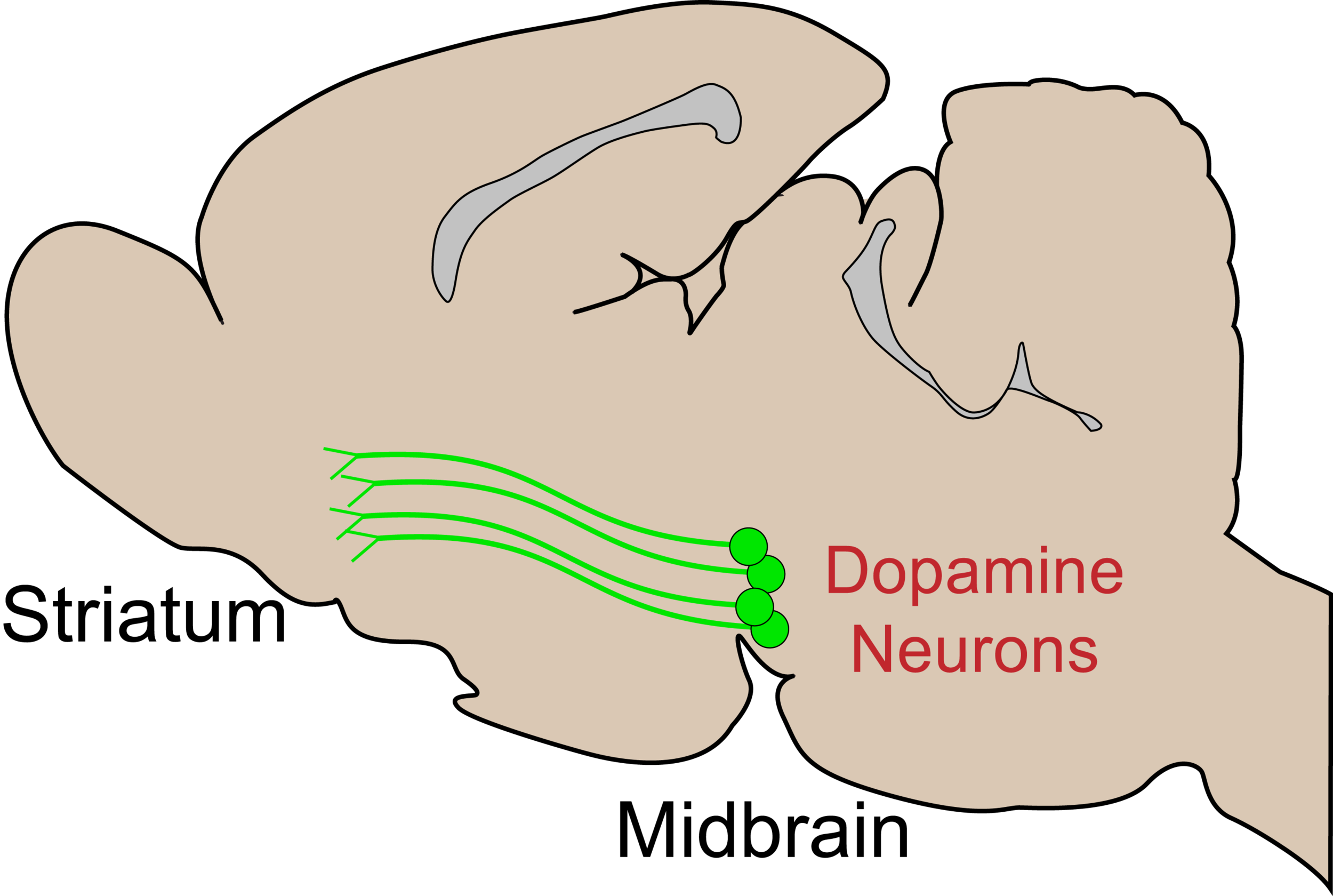
Neurons releasing dopamine arise from deep in the brain and project to a variety of regions, including the striatum, where their signaling influences reward-related processes. Shown is a cartoon of dopamine neurons in the rat brain, but the primary circuits are similar in humans.
Signaling from dopamine neurons contributes broadly to reward processes, but their function in anatomically-defined circuits is not well understood. A major focus of my research is to determine how subpopulations of dopamine neurons contribute to distinct facets of reward.
To this end, I use genetic-targeting strategies to manipulate and record activity from dopamine neurons as rats engage in behavior.

Heterogeneous cue-evoked motivational signals from distinct dopamine (DA) neuron projections. Ventral striatal dopamine neurons promote "attraction" to conditioned cues (CS), while dorsal striatal dopamine neurons promote movement. Dopamine neuron terminals in the striatum are visualized here in green.
Saunders et al., in prep (2016 SFN Poster).
A major way dopamine neurons influence reward seeking is by modulating the value of environmental stimuli ("cues") that become associated with rewarding outcomes. Exactly how this process occurs at a neural-circuit level remains unclear. In a Pavlovian conditioning task, I found that a cue paired with optogenetic activation of dopamine neurons acquired the ability to evoke behaviors on its own, even though it was never associated with "real" reward (i.e., food or water). The topography of cue-evoked behaviors, however, differed according to the dopamine neuron subpopulation engaged. This project demonstrates that dopamine neuron activity can take the place of a natural rewards to imbue environmental cues with motivational value to drive various behaviors.

Fiber photometry allows for the measurement of calcium signaling, as a proxy for neural activity, via genetically-encoded calcium indicators (GCaMP6). Here photometry was combined with optogenetic manipulation of the same neurons, during Pavlovian conditioning. Saunders et al., in prep.
New technologies make it possible to simultaneously manipulate and measure activity from neurons based on genetic and/or anatomical identity. In these studies I use fiber photometry to record calcium activity (via GCaMP6) in dopamine neurons. This, coupled with optogenetic manipulations (via ChrimsonR), allows for assessment of cue-evoked activity in dopamine neurons during Pavlovian conditioning.
Midbrain circuit dynamics in cocaine seeking

Imaging calcium fluorescence using miniature microscopes makes it possible to assess single neuron activity in freely moving animals. Shown is an unprocessed movie of GABAergic neurons in the midbrain of a mouse engaged in cocaine-related behaviors.

Current technology requires tethering to a data acquisition source, but new microscopes are under development that will allow for wireless recordings, which I will use in the future for imaging in rats.
As part of a pending K99/R00 grant from the National Institute on Drug Abuse, I am using head-mounted miniature microscopes to image calcium activity with single cell resolution in midbrain circuits. This project will identify how genetically (dopaminergic, GABAergic) and projection (striatal, thalamic) defined midbrain neurons respond to cocaine exposure, how they change with repeated drug experience, and how they are engaged by drug-associated cues to promote or suppress drug-seeking.
Future Directions
General Aim: Characterize serial mesostriatal circuit dynamics across cue conditioning. This project will utilize multi-site fiber photometry, optogenetic, and genetic deletion strategies in dopamine neurons to investigate the hypothesis that a progressive recruitment of ventral-to-dorsal projecting dopamine neuron signaling underlies motivational shifts in Pavlovian cue-evoked behavior.

Here I will condition cues to optogenetic VTA DA stimulation and track cue-evoked calcium activity in axons in the nucleus accumbens and dorsolateral striatum across training.
General Aim: Characterize midbrain neuronal ensembles underlying distinct Pavlovian conditioned responses. This project will use in vivo calcium imaging to investigate the hypothesis that distinct dopamine neuronal ensembles in the ventral tegmental area encode conditioned responses that reflect cue attraction "wanting" versus cue-evoked movement invigoration.
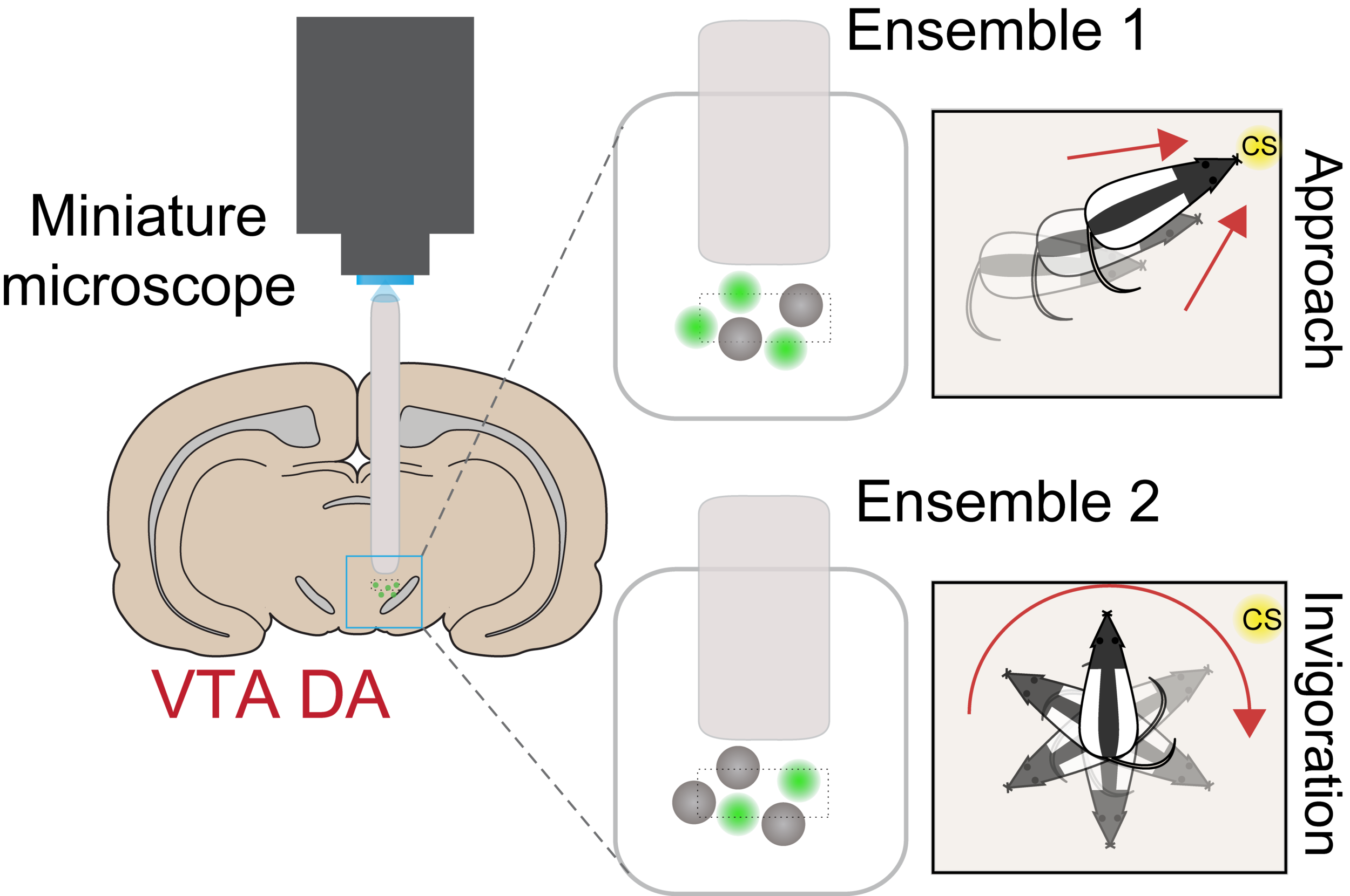
Heterogeneous DA neuron activity will be further explored with projection-defined imaging studies.